


Degree in Radio Imaging Technician Course- What All the Course Covers?

February 04, 2025

Are you looking forward to a career in radio imaging in the healthcare sector? Are you perplexed by the information available online? Aren’t you able to choose the course and college? Do you want to know about the degree in radio imaging technician course? If yes is what you might answer, reading further will help you understand this course and the topics it covers. So, here we go.
What is the Radio Imaging Technician Course?
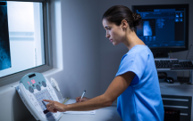
Medical radio imaging technology means the use of several imaging methods in the healthcare field to generate images of the human body parts for treatment and diagnostic uses. A degree course in this field covers several imaging modalities like Positron Emission Tomography (PET), X-ray, Magnetic Resonance Imaging (MRI), Angiography, and Computed Tomography (CT scan), among others. This job prepares aspiring candidates for a promising profession as a radio imaging technician.
Here are the topics that the course covers over three years.
- Image interpretation that includes MRI machines, X-ray machines, and CT scanners.
- The principles of radiation physics and medical imaging.
- Image analysis and processing that includes computer-aided diagnosis and digital image processing.
- Radiation safety in the three-year course teaches about the safe and careful usage of ionizing radiation in patient protection and medical imaging.
- Techniques of image acquisition, including exposure parameters and positioning of patients.
Aspirants who become graduates in this field can fetch a job as medical sonographers, medical radiologic technologists, and medical radiation professionals.
Radio Imaging Technology Scope and Details
The scope of this course is immense. It covers a range of therapeutic and diagnostic procedures that make use of several imaging methods to produce different images of the human body parts. Listed below are some of the main areas of focus and expertise within this field.
Magnetic Resonance Imaging – Commonly known as MRI, it uses radio waves and a magnetic field to generate images of the internal structure of the human body.
Interventional Radiology—This is an important subspecialty of the radiology imaging field that makes use of imaging guidance to perform various minimally invasive procedures like drainage of fluid collections, biopsies, and embolization.
Computed Tomography—Also known as CT scans, it uses computer processing and X-rays to generate detailed and cross-sectional human body images.
X-ray Imaging—This is one of the most widely used imaging modalities that produces images of internal organs and bones using ionizing radiation.
Ultrasound Imaging—This is a known non-invasive imaging method using high-frequency sound waves for producing images of tissues and internal organs.
Nuclear Medicine—This is a crucial part of the course wherein candidates learn about the use of radioactive materials to generate clear images of the body’s tissues and organs and understand and assess their function.
Positron Emission Tomography—Also known as PET scans, it relies on radioactive tracers to produce sharp and clear images of metabolic processes within the human body. With the advancement in the healthcare sector, the scope of radio imaging technology expands and evolves, paving the way for promising career opportunities for radio imaging technicians. Enrolling in a bachelor in radio imaging technician course ensures a promising career in this field. Let your career in the healthcare sector grow by becoming a radio imaging technician.

